
추천 제품


Grade
Molecular Biology
Quality Level
양식
liquid
농도
1.25 ×
기술
electrophoresis: suitable
외래 활성
RNase, none detected
저장 온도
−20°C
−20°C
유사한 제품을 찾으십니까? 방문 제품 비교 안내
일반 설명
RNA loading buffer is used as a tracking dye during RNA electrophoresis. The RNA loading dye has a slight negative charge and will migrate the same direction as RNA, allowing the user to monitor the progress of molecules moving through the gel. The rate of migration varies with gel composition. Dilute 1:3 to 1:6 with sample, heat to 65C for ten minutes and chill on ice before loading.
애플리케이션
RNA sample loading buffer is especially formulated for electrophoresis of RNA on formaldehyde-agarose gels with or without ethidium bromide. Ethidium bromide is not recommended for gel staining prior to Northern blot detection because the presence of ethidium bromide in agarose gels or the loading buffer can cause poor transfer efficiency.
Suitable for use with formaldehyde-agarose gels used in Northern blotting procedures.
RNA Sample Loading Buffer has been used as a sample loading buffer in northern blot.
RNA Sample Loading Buffer has been used as a sample loading buffer in northern blot.
성분
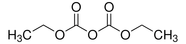
Deionized formamide 62.5% (v/v), formaldehyde 1.14 M, bromphenol blue 200 μg/mL, xylene cyanole 200 μg/mL, MOPS-EDTA-sodium acetate at 1.25× working concentration.
수량
Recommended usage: Add 1 volume sample to 2-5 volumes of sample loading buffer and mix well. The sample should be heated to 65 °C for 10 minutes, and then chilled on ice immediately before loading on the gel.
제조 메모
Deionized formamide 62.5% (v/v), formaldehyde 1.14 M, bromphenol blue 200 μg/mL, xylene cyanole 200 μg/mL, MOPS-EDTA-sodium acetate at 1.25× working concentration.
관련 제품
제품 번호
설명
가격


신호어
Danger
유해 및 위험 성명서
Hazard Classifications
Acute Tox. 4 Inhalation - Carc. 1B - Muta. 2 - Repr. 1B - Skin Sens. 1 - STOT RE 2 Oral
표적 기관
Blood
Storage Class Code
6.1C - Combustible, acute toxic Cat.3 / toxic compounds or compounds which causing chronic effects
WGK
WGK 3
Flash Point (°F)
Not applicable
Flash Point (°C)
Not applicable
이미 열람한 고객


Dennis L Stevens et al.
The Journal of infectious diseases, 195(2), 202-211 (2006-12-28)
Extracellular protein toxins contribute to the pathogenesis of a wide variety of Staphylococcus aureus infections. The present study investigated the effects that cell-wall active antibiotics and protein-synthesis inhibitors have on transcription and translation of genes for Panton-Valentine leukocidin, alpha-hemolysin, and
Impact of antibiotics on expression of virulence-associated exotoxin genes in methicillin-sensitive and methicillin-resistant Staphylococcus aureus.
Stevens DL, et al.
The Journal of Infectious Diseases, 195, 202-211 (2007)
Erika C von Grote et al.
Molecular bioSystems, 7(1), 150-161 (2010-08-24)
Cytokines are important mediators of the wound healing response. However, sampling of cytokines from the interstitial fluid at a healing wound site in experimental animals is a challenge. Microdialysis sampling is an in vivo collection option for this purpose as
The effect of massive small bowel resection and oral epidermal growth factor therapy on SGLT-1 distribution in rabbit distal remnant.
Chung BM, et al.
Pediatric Research, 55, 19-26 (2004)
Amy E Bryant et al.
Journal of medical microbiology, 68(3), 456-466 (2019-01-25)
Extracellular protein toxins contribute to the pathogenesis of Staphylococcus aureus infections. The present study compared the effects of iclaprim and trimethoprim - two folic acid synthesis inhibitors - with nafcillin and vancomycin on production of Panton-Valentine leukocidin (PVL), alpha haemolysin
자사의 과학자팀은 생명 과학, 재료 과학, 화학 합성, 크로마토그래피, 분석 및 기타 많은 영역을 포함한 모든 과학 분야에 경험이 있습니다..
고객지원팀으로 연락바랍니다.