
추천 제품


생물학적 소스
rabbit
Quality Level
결합
unconjugated
항체 형태
affinity isolated antibody
항체 생산 유형
primary antibodies
클론
polyclonal
제품 라인
Prestige Antibodies® Powered by Atlas Antibodies
양식
buffered aqueous glycerol solution
종 반응성
human
향상된 검증
orthogonal RNAseq
independent
Learn more about Antibody Enhanced Validation
기술
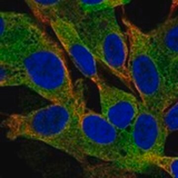
immunofluorescence: 0.25-2 μg/mL
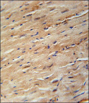
immunohistochemistry: 1:50-1:200
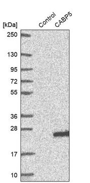
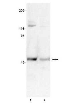
western blot: 0.04-0.4 μg/mL
면역원 서열
VKRLEKQNLEKDQVNKDLTEKLEALESLRLQEQAALETEDGEGLQQTLRDLAQAVLSDSESGVQLSGSERTADASNGS
UniProt 수납 번호
배송 상태
wet ice
저장 온도
−20°C
타겟 번역 후 변형
unmodified
유전자 정보
human ... CROCC(9696)
일반 설명
The gene CROCC (ciliary rootlet coiled-coil protein) is mapped to human chromosome 1p36.13. CROCC is commonly referred to as rootletin.
면역원
Rootletin recombinant protein epitope signature tag (PrEST)
애플리케이션
Anti-CROCC antibody produced in rabbit, a Prestige Antibody, is developed and validated by the Human Protein Atlas (HPA) project . Each antibody is tested by immunohistochemistry against hundreds of normal and disease tissues. These images can be viewed on the Human Protein Atlas (HPA) site by clicking on the Image Gallery link. The antibodies are also tested using immunofluorescence and western blotting. To view these protocols and other useful information about Prestige Antibodies and the HPA, visit sigma.com/prestige.
생화학적/생리학적 작용
CROCC (ciliary rootlet coiled-coil protein) is important for maintaining centrosome cohesion. It is responsible for forming centriole-associated fibers. CROCC also functions as a ciliary rootlet structural component. Down-regulation of CROCC in mice results in photoreceptor degeneration and negatively affects mucociliary clearance.
특징 및 장점
Prestige Antibodies® are highly characterized and extensively validated antibodies with the added benefit of all available characterization data for each target being accessible via the Human Protein Atlas portal linked just below the product name at the top of this page. The uniqueness and low cross-reactivity of the Prestige Antibodies® to other proteins are due to a thorough selection of antigen regions, affinity purification, and stringent selection. Prestige antigen controls are available for every corresponding Prestige Antibody and can be found in the linkage section.
Every Prestige Antibody is tested in the following ways:
Every Prestige Antibody is tested in the following ways:
- IHC tissue array of 44 normal human tissues and 20 of the most common cancer type tissues.
- Protein array of 364 human recombinant protein fragments.
결합
Corresponding Antigen APREST76003
물리적 형태
Solution in phosphate-buffered saline, pH 7.2, containing 40% glycerol and 0.02% sodium azide
법적 정보
Prestige Antibodies is a registered trademark of Merck KGaA, Darmstadt, Germany
면책조항
Unless otherwise stated in our catalog or other company documentation accompanying the product(s), our products are intended for research use only and are not to be used for any other purpose, which includes but is not limited to, unauthorized commercial uses, in vitro diagnostic uses, ex vivo or in vivo therapeutic uses or any type of consumption or application to humans or animals.
적합한 제품을 찾을 수 없으신가요?
당사의 제품 선택기 도구.을(를) 시도해 보세요.
Storage Class Code
10 - Combustible liquids
WGK
WGK 1
Flash Point (°F)
Not applicable
Flash Point (°C)
Not applicable
가장 최신 버전 중 하나를 선택하세요:
Linda Shyue Huey Chuang et al.
Cell cycle (Georgetown, Tex.), 11(10), 1938-1947 (2012-05-01)
RUNX family proteins are critical regulators of lineage differentiation during development. The high prevalence of RUNX mutation/epigenetic inactivation in human cancer indicates a causative role for dysfunctional RUNX in carcinogenesis. This is supported by well-documented evidence of functional interaction of
Rosangela Artuso et al.
Journal of human genetics, 56(7), 508-515 (2011-05-20)
MECP2 mutations are responsible for two different phenotypes in females, classical Rett syndrome and the milder Zappella variant (Z-RTT). We investigated whether copy number variants (CNVs) may modulate the phenotype by comparison of array-CGH data from two discordant pairs of
DNA damage-induced centrosome amplification occurs via excessive formation of centriolar satellites.
H Löffler et al.
Oncogene, 32(24), 2963-2972 (2012-07-25)
Centrosome amplification is a frequent phenomenon in malignancies and may facilitate tumorigenesis by promoting chromosomal instability. On the other hand, a centrosome inactivation checkpoint comprising centrosome amplification leading to elimination of cells by mitotic catastrophe has been described in response
Julia Wallmeier et al.
Nature genetics, 46(6), 646-651 (2014-04-22)
Using a whole-exome sequencing strategy, we identified recessive CCNO (encoding cyclin O) mutations in 16 individuals suffering from chronic destructive lung disease due to insufficient airway clearance. Respiratory epithelial cells showed a marked reduction in the number of multiple motile
Susanne Bahe et al.
The Journal of cell biology, 171(1), 27-33 (2005-10-06)
After duplication of the centriole pair during S phase, the centrosome functions as a single microtubule-organizing center until the onset of mitosis, when the duplicated centrosomes separate for bipolar spindle formation. The mechanisms regulating centrosome cohesion and separation during the
자사의 과학자팀은 생명 과학, 재료 과학, 화학 합성, 크로마토그래피, 분석 및 기타 많은 영역을 포함한 모든 과학 분야에 경험이 있습니다..
고객지원팀으로 연락바랍니다.