
추천 제품


생물학적 소스
mouse
Quality Level
항체 형태
purified immunoglobulin
항체 생산 유형
primary antibodies
클론
A60, monoclonal
종 반응성
avian, pig, chicken, human, rat, salamander, ferret, mouse
제조업체/상표
Chemicon®
기술
flow cytometry: suitable
immunocytochemistry: suitable
immunofluorescence: suitable
immunohistochemistry (formalin-fixed, paraffin-embedded sections): suitable
immunoprecipitation (IP): suitable
western blot: suitable
동형
IgG1
배송 상태
wet ice
타겟 번역 후 변형
unmodified
유전자 정보
human ... RBFOX3(146713)
mouse ... Rbfox3(52897)
rat ... Rbfox3(287847)
일반 설명
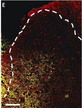
Anti-NeuN antibody (NEUronal Nuclei; clone A60) specifically recognizes the DNA-binding, neuron-specific protein NeuN, which is present in most CNS and PNS neuronal cell types of all vertebrates tested. NeuN protein distributions are apparently restricted to neuronal nuclei, perikarya and some proximal neuronal processes in both fetal and adult brain although, some neurons fail to be recognized by NeuN at all ages: INL retinal cells, Cajal-Retzius cells, Purkinje cells, inferior olivary and dentate nucleus neurons, and sympathetic ganglion cells are examples (Mullen et al., 1992; Wolf et al., 1996). Immunohistochemically detectable NeuN protein first appears at developmental timepoints that correspond with the withdrawal of the neuron from the cell cycle and/or with the initiation of terminal differentiation of the neuron (Mullen et al., 1992). Immunoreactivity appears around E9.5 in the mouse neural tube and is extensive throughout the developing nervous system by E12.5. Strong nuclear staining suggests a nuclear regulatory protein function; however, no evidence currently exists as to whether the NeuN protein antigen has a function in the distal cytoplasm or whether it is merely synthesized there before being transported back into the nucleus. No difference between protein isolated from purified nuclei and whole brain extract on immunoblots has been found (Mullen et al., 1992).
특이성
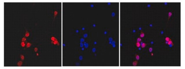
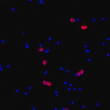
MILLIPORE′s exclusive monoclonal antibody to vertebrate neuron-specific nuclear protein called NeuN (or Neuronal Nuclei) reacts with most neuronal cell types throughout the nervous system of mice including cerebellum, cerebral cortex, hippocampus, thalamus, spinal cord and neurons in the peripheral nervous system including dorsal root ganglia, sympathetic chain ganglia and enteric ganglia. Developmentally, immunoreactivity is first observed shortly after neurons have become postmitotic, no staining has been observed in proliferative zones. The immunohistochemical staining is primarily localized in the nucleus of the neurons with lighter staining in the cytoplasm. The few cell types not reactive with MAB377 include Purkinje, mitral and photoreceptor cells. The antibody is an excellent marker for neurons in primary cultures and in retinoic acid-stimulated P19 cells. It is also useful for identifying neurons in transplants.
면역원
Purified cell nuclei from mouse brain
애플리케이션
Anti-NeuN Antibody, clone A60 detects level of NeuN and has been published and validated for use in FC, IC, IF, IH, IH(P), IP and WB.
Research Category
Neuroscience
Neuroscience
Research Sub Category
Neuronal & Glial Markers
Neuronal & Glial Markers
Western Blot Analysis:
A previous lot of this antibody recognized 2-3 bands in the 46-48 kDa range and possibly another band at approximately 66 kDa.
Immunocytochemistry:
1:10-1:100 dilution from a previous lot was used. Neurons in culture should be permeablized with 0.1% triton X-100. All primary antibody dilutions should be performed with simple solutions containing only buffer and primary antibody without excess protein blocks or detergents.
Immunohistochemistry:
1:100-1:1,000. The antibody works best on polyester wax embedded tissue but also works on paraffin embedded tissue at a lower working dilution. The antibody works well with formaldehyde-based fixatives. Citric acid and microwave pretreatment has been used successfully (Sarnat, 1998).
Immunohistochemistry(paraffin) Analysis: A previous lot was used for IH(P).
Optimal working dilutions must be determined by end user.
품질
Routinely evaluated by immunohistochemistry on brain tissue.
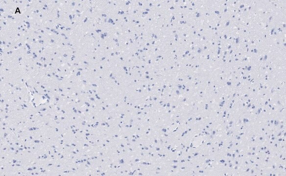
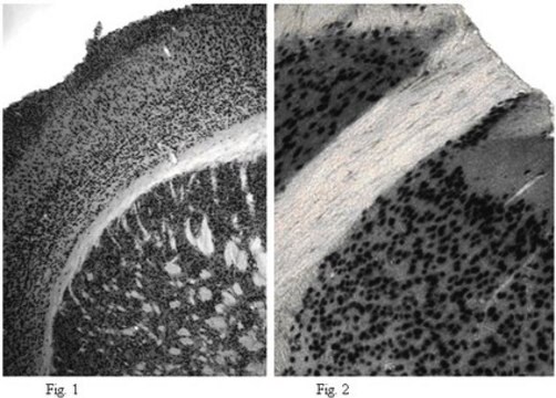
Immunohistochemistry(paraffin) Analysis:
NeuN (cat. # MAB377) staining pattern/morphology in rat cerebellum. Tissue pretreated with Citrate, pH 6.0. This lot of antibody was diluted to 1:100, using IHC-Select Detection with HRP-DAB. Immunoreactivity is seen as nuclear staining in the neurons in the granular layer. Note that there is no signal detected in the nucleus of Purkinje cells.
Optimal Staining With Citrate Buffer, pH 6.0, Epitope Retrieval: Rat Cerebellum
표적 설명
46/48 kDa
물리적 형태
Format: Purified
Protein A purified
Purified mouse immunoglobulin IgG1 liquid in buffer containing 0.02 M phosphate buffer, 0.25 M NaCl, pH 7.6 with 0.1% sodium azide.
저장 및 안정성
Stable for 6 months at 2-8ºC from date of receipt.
분석 메모
Control
Positive control -Brain Tissue. Negative control - Any non neuronal tissue eg Fibroblasts
Positive control -Brain Tissue. Negative control - Any non neuronal tissue eg Fibroblasts
법적 정보
CHEMICON is a registered trademark of Merck KGaA, Darmstadt, Germany
면책조항
Unless otherwise stated in our catalog or other company documentation accompanying the product(s), our products are intended for research use only and are not to be used for any other purpose, which includes but is not limited to, unauthorized commercial uses, in vitro diagnostic uses, ex vivo or in vivo therapeutic uses or any type of consumption or application to humans or animals.
적합한 제품을 찾을 수 없으신가요?
당사의 제품 선택기 도구.을(를) 시도해 보세요.
Storage Class Code
12 - Non Combustible Liquids
WGK
WGK 2
Flash Point (°F)
Not applicable
Flash Point (°C)
Not applicable
시험 성적서(COA)
제품의 로트/배치 번호를 입력하여 시험 성적서(COA)을 검색하십시오. 로트 및 배치 번호는 제품 라벨에 있는 ‘로트’ 또는 ‘배치’라는 용어 뒤에서 찾을 수 있습니다.
이미 열람한 고객


Presubiculum stimulation in vivo evokes distinct oscillations in superficial and deep entorhinal cortex layers in chronic epileptic rats.
Tolner, EA; Kloosterman, F; van Vliet, EA; Witter, MP; Silva, FH; Gorter, JA
The Journal of Neuroscience null
A U Zaidi et al.
The journal of histochemistry and cytochemistry : official journal of the Histochemistry Society, 48(10), 1369-1375 (2000-09-16)
To understand the biological relationships among various molecules, it is necessary to define the cellular expression patterns of multiple genes and gene products. Relatively simple methods for performing multi-label immunohistochemical detection are available. However, there is a paucity of techniques
Isotropic fractionator: a simple, rapid method for the quantification of total cell and neuron numbers in the brain.
Herculano-Houzel, Suzana and Lent, Roberto
The Journal of Neuroscience, 25, 2518-2521 (2005)
A role for prefrontal cortex in memory storage for trace fear conditioning.
Runyan, Jason D, et al.
The Journal of Neuroscience, 24, 1288-1295 (2004)
Different expression patterns of Bcl-2, Bcl-xl, and Bax proteins after sublethal forebrain ischemia in C57Black/Crj6 mouse striatum.
Wu, C; Fujihara, H; Yao, J; Qi, S; Li, H; Shimoji, K; Baba, H
Stroke null
자사의 과학자팀은 생명 과학, 재료 과학, 화학 합성, 크로마토그래피, 분석 및 기타 많은 영역을 포함한 모든 과학 분야에 경험이 있습니다..
고객지원팀으로 연락바랍니다.