Accéder au contenu
MilliporeSigma
Toutes les photos(1)

Principaux documents

MABS2269

Sigma-Aldrich

Anti-phospho-PD-1 (Tyr248) Antibody, clone 407.6G12

new

Synonyme(s) :

CD279, Programmed cell death protein 1, Protein PD-1, hPD-1

Se connecterpour consulter vos tarifs contractuels et ceux de votre entreprise/organisme


About This Item

Code UNSPSC :
12352203

Source biologique

mouse

Niveau de qualité

Forme d'anticorps

purified antibody

Type de produit anticorps

primary antibodies

Clone

407.6G12, monoclonal

Poids mol.

calculated mol wt 31.65 kDa
observed mol wt ~55 kDa

Produit purifié par

using protein G

Espèces réactives

mouse, human

Réactivité de l'espèce (prédite par homologie)

monkey

Conditionnement

antibody small pack of 100

Technique(s)

flow cytometry: suitable
western blot: suitable

Isotype

IgG2aκ

Séquence de l'épitope

C-terminal half

Numéro d'accès Protein ID

Numéro d'accès UniProt

Température de stockage

2-8°C

Informations sur le gène

human ... PDCD1(5133)

Spécificité

Clone 407.6G12 is a mouse monoclonal antibody that specifically detects PD-1 (CD279) phosphorylated on tyrosine 248.

Immunogène

KLH-conjugated linear peptide corresponding to 13 amino acids surrounding phospho-tyrosine 248 from the C-terminal half of human PD-1 (CD279).

Application

Quality Control Testing

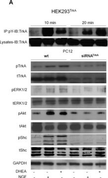
Evaluated by Western Blotting in lysate from Jurkat cells treated with pervanadate (0.1 mM; 5 min.at 37C).

Western Blotting Analysis (WB): A 1:1000 dilution of this antibody detected PD-1 phosphorylated on tyrosine 248 in lysate from Jurkat cells treated with pervanadate (0.1 mM; 5 min. at 37C), but not in lysate from untreated Jurkat cells.

Tested Applications

Western Blotting Analysis: A representative lot detected PD-1 (CD279) in Western Blotting application (Bu, X., et al. (2021). Cancer Immunol Res. 9(12):1465-1475).

Flow Cytometry Analysis: A representative lot detected PD-1 (CD279) in Flow Cytometry application (Bu, X., et al. (2021). Cancer Immunol Res. 9(12):1465-1475).

Note: Actual optimal working dilutions must be determined by end user as specimens, and experimental conditions may vary with the end user.

Description de la cible

Programmed cell death protein 1 (UniProt: Q15116; also known as Protein PD-1, hPD-1, CD279) is encoded by the PDCD1 (also known as PD1) gene (Gene ID: 5133) in human. PD-1 is a monomeric inhibitory cell surface receptor involved in the regulation of T-cell function during immunity and tolerance. PD-1 and its ligands, PD-L1 and PD-L2 play a key role in the maintenance of peripheral tolerance, a process by which the quiescence of autoreactive mature T cells is maintained. PD-1 is a single-pass type I membrane protein, synthesized with a signal peptide (aa 1-20), which is subsequently cleaved off. The mature form contains an extracellular domain (aa 24-170), a transmembrane domain (aa 171-191), and a cytoplasmic domain (aa 192-288). PD-1 also contains a single N-terminal immunoglobulin variable region (IgV) like domain, an ITIM motif (aa 221-226) and an ITSM motif ( aa 247-251). Tyrosine 248 falls in ITSM motif and located within the intracellular domain of PD-1 and, phosphorylation of PD-1 at Tyrosine 248 is a significant post-translational modification that influences its function. Src family kinases, such as Lck and Fyn, are involved in the initial phosphorylation of PD-1. Upon binding of PD-1 to its ligands (PD-L1 or PD-L2), phosphorylation at Tyr248 triggers downstream signaling pathways. PD-1 phosphorylation at Tyr248 promotes the recruitment of the protein tyrosine phosphatase PTPN11/SHP-2 that mediates de-phosphorylation of key TCR proximal signaling molecules, such as ZAP70, PRKCQ/PKC theta and CD247/CD3zeta. The phosphorylation at Tyr248 is crucial for the full inhibitory effect of PD-1 on T-cell activity. It contributes to the suppression of T-cell proliferation, cytokine production, and cytotoxic activity. Studies demonstrate that phosphorylation of the ITSM motif of PD-1 marks dysfunctional T cells that may be rescued with PD-1 blockade. Detection of phospho-PD-1 in TILs is a potential biomarker for PD-1 immunotherapy responses. (Ref.: Bu, X., et al. (2021). Cancer Immunol Res. 9(12):1465-1475).

Forme physique

Purified mouse monoclonal antibody IgG2a in buffer containing 0.1 M Tris-Glycine (pH 7.4), 150 mM NaCl with 0.05% sodium azide.

Reconstitution

1.0 mg/mL. Please refer to guidance on suggested starting dilutions and/or titers per application and sample type.

Stockage et stabilité

Recommended storage: +2°C to +8°C.

Autres remarques

Concentration: Please refer to the Certificate of Analysis for the lot-specific concentration.

Clause de non-responsabilité

Unless otherwise stated in our catalog or other company documentation accompanying the product(s), our products are intended for research use only and are not to be used for any other purpose, which includes but is not limited to, unauthorized commercial uses, in vitro diagnostic uses, ex vivo or in vivo therapeutic uses or any type of consumption or application to humans or animals.

Vous ne trouvez pas le bon produit ?  

Essayez notre Outil de sélection de produits.

Code de la classe de stockage

12 - Non Combustible Liquids

Classe de danger pour l'eau (WGK)

WGK 1

Point d'éclair (°F)

Not applicable

Point d'éclair (°C)

Not applicable


Certificats d'analyse (COA)

Recherchez un Certificats d'analyse (COA) en saisissant le numéro de lot du produit. Les numéros de lot figurent sur l'étiquette du produit après les mots "Lot" ou "Batch".

Déjà en possession de ce produit ?

Retrouvez la documentation relative aux produits que vous avez récemment achetés dans la Bibliothèque de documents.

Consulter la Bibliothèque de documents

Notre équipe de scientifiques dispose d'une expérience dans tous les secteurs de la recherche, notamment en sciences de la vie, science des matériaux, synthèse chimique, chromatographie, analyse et dans de nombreux autres domaines..

Contacter notre Service technique