
MABN798
Anti-retSDR1 Antibody, clone A11
clone A11, from mouse
Synonyme(s) :
Short-chain dehydrogenase/reductase 3, DD83.1, Retinol dehydrogenase 17, Retinal short-chain dehydrogenase/reductase 1, retSDR1, Short chain dehydrogenase/reductase family 16C member 1
About This Item
Produits recommandés


Source biologique
mouse
Niveau de qualité
Forme d'anticorps
purified immunoglobulin
Type de produit anticorps
primary antibodies
Clone
A11, monoclonal
Espèces réactives
human, bovine, rat
Technique(s)
immunofluorescence: suitable
immunohistochemistry: suitable (paraffin)
western blot: suitable
Isotype
IgG1κ
Numéro d'accès NCBI
Numéro d'accès UniProt
Modification post-traductionnelle de la cible
unmodified
Informations sur le gène
human ... DHRS3(9249)
Description générale
Spécificité
Immunogène
Application
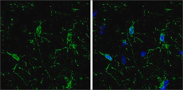
Immunofluorescence Analysis: Clone A11 hybridoma culture supernatant immunostained cone outer segments and a subset of inner nuclear layer somata in 4% paraformaldeyde-fixed, agarose-embedded bovine retina sections by fluorescent confocal microscopy (Courtesy of Francoise Haeseleer, Ph.D., University of Washington, Seattle, WA).
Western Blotting Analysis: A representative lot detected a greatly upregulated retSDR1 protein expression following p53 or TAp63γ overexpression induction in DLD-1 human colorectal adenocarcinoma cells (Kirschner, R.D., et al. (2010). Cell Cycle. 9(11):2177-2188).
Immunofluorescence Analysis: A representative lot immunostained cone, but not rod, outer segments in 4% paraformaldeyde-fixed, agarose-embedded bovine retina sections by fluorescent confocal microscopy. Immunoreactivity was also observed in a subset of somata localized in the inner nuclear layer (Haeseleer, F., et al. (1998). J. Biol. Chem. 273(34):21790-21799).
Immunohistochemistry Analysis: A representative lot immunostained cone, but not rod, outer segments in 4% paraformaldeyde-fixed bovine retina whole mount sections. Immunoreactivity was also observed in a subset of somata localized in the inner nuclear layer (Haeseleer, F., et al. (1998). J. Biol. Chem. 273(34):21790-21799).
Neuroscience
Sensory & PNS
Qualité
Western Blotting Analysis: 0.5 µg/mL of this antibody detected the expression of full-length human retSDR1 in 10 µg of lysate from transfected, but not untransfected HEK293 cells.
Description de la cible
Forme physique
Stockage et stabilité
Autres remarques
Clause de non-responsabilité
Vous ne trouvez pas le bon produit ?
Essayez notre Outil de sélection de produits.
Code de la classe de stockage
12 - Non Combustible Liquids
Classe de danger pour l'eau (WGK)
WGK 1
Point d'éclair (°F)
Not applicable
Point d'éclair (°C)
Not applicable
Certificats d'analyse (COA)
Recherchez un Certificats d'analyse (COA) en saisissant le numéro de lot du produit. Les numéros de lot figurent sur l'étiquette du produit après les mots "Lot" ou "Batch".
Déjà en possession de ce produit ?
Retrouvez la documentation relative aux produits que vous avez récemment achetés dans la Bibliothèque de documents.
Notre équipe de scientifiques dispose d'une expérience dans tous les secteurs de la recherche, notamment en sciences de la vie, science des matériaux, synthèse chimique, chromatographie, analyse et dans de nombreux autres domaines..
Contacter notre Service technique