
MABS1981
Anti-TfR2 Antibody, clone 9F8-1C11
clone 9F8 1C11, from mouse
Synonym(s):
Transferrin receptor protein 2
Sign Into View Organizational & Contract Pricing
Select a Size
All Photos(1)
Select a Size
Change View
About This Item
UNSPSC Code:
12352203
eCl@ss:
32160702
NACRES:
NA.43
Recommended Products


biological source
mouse
Quality Level
antibody form
purified immunoglobulin
antibody product type
primary antibodies
clone
9F8 1C11, monoclonal
species reactivity
human
technique(s)
immunocytochemistry: suitable
immunoprecipitation (IP): suitable
western blot: suitable
isotype
IgG1κ
NCBI accession no.
UniProt accession no.
General description
Transferrin receptor protein 2 (UniProt: Q9UP52; also known as TfR2) is encoded by the TFR2 gene (Gene ID: 70360 in human. TfR2 is a single-pass type II membrane glycoprotein that plays a critical role in iron metabolism. It has a large C-terminal ectodomain and a small N-terminal cytoplasmic domain and shares 45% amino acid sequence with extracellular region of transferrin receptor (TfR). This homodimeric membrane receptor binds 2 molecules of transferrin and is internalized into endosomes that are acidified, resulting in the release of iron from transferrin. TfR2 contains a cytoplasmic internalization motif similar to TfR, and has 2 cysteines, which form inter subunit disulfide bonds, in the ectodomain proximal to the transmembrane domain. TfR2 mediates cellular uptake of transferrin-bound iron in a non-iron dependent manner. Both TfR and TfR2 bind diferric transferrin better than apo-transferrin at neutral pH. However, TFR2 displays about 25-fold lower affinity for transferrin compared to TfR and its expression is not controlled by cellular levels of iron. TfR2 is also suggested to be involved in involved in hepatocyte function and erythrocyte differentiation. Mutations in TFR2 gene are known to cause hemochromatosis 3 that is characterized by an iron overload where excess iron is deposited in several organs leading to organ failure. (Ref.: Kawabata H., et al (1999). J. Biol. Chem. 274 (30): 20826-20832; Vogt, TM et al. (2003). Blood 101(5):2008-2014.
Specificity
Clone 9F8-1C11 sprcifically detects human TfR2 and does exhibit reactivity with TfR.
Immunogen
Epitope: extracellular domain
Purified ectodomains of the human TfR2.
Application
Anti-TfR2, clone 9F8-1C11 Antibody, Cat. No. MABS1981, is a highly specific mouse monoclonal antibody, that targets Tfr2 and has been tested in Western Blotting, Immunoprecipitation, and Immunocytochemistry.
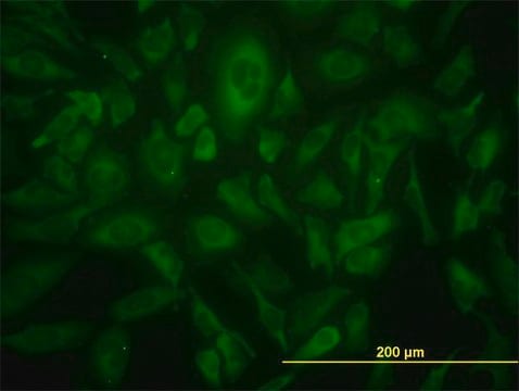
Immunocytochemistry Analysis: A representative lot detected Tfr2 in Immunocytochmeistry applications (Vogt, T.M., et. al. (2003). Blood. 101(5):2008-14; Zhao, N., et. al. (2013). Biochemistry. 52(19):3310-9).
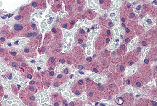
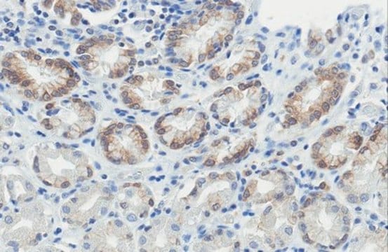
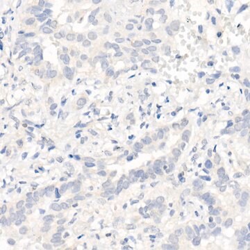
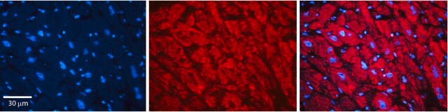
Immunohistochemistry Analysis: A representative lot detected Tfr2 in Immunohistochemistry applications (Vogt, T.M., et. al. (2003). Blood. 101(5):2008-14).
Western Blotting Analysis: A representative lot detected Tfr2 in Western Blotting applications (Vogt, T.M., et. al. (2003). Blood. 101(5):2008-14; Byrne, S.L., et. al. (2013). PLoS One. 8(7):e70199; Zhao, N., et. al. (2013). Biochemistry. 52(19):3310-9).
Immunoprecipitation Analysis: A representative lot detected Tfr2 in Immunoprecipitation applications (Vogt, T.M., et. al. (2003). Blood. 101(5):2008-14; Johnson, M.B., et. al. (2004). Blood. 104(13):4287-93).
Research Category
Epigenetics & Nuclear Function
Epigenetics & Nuclear Function
Quality
Evaluated by Western Blotting in K562 cell lysate.
Western Blotting Analysis: 1 µg/mL of this antibody detected Tfr2 in 10 µg of K562 cell lysaste.
Western Blotting Analysis: 1 µg/mL of this antibody detected Tfr2 in 10 µg of K562 cell lysaste.
Target description
~108 kDa observed; 88.76 kDa calculated. Uncharacterized bands may be observed in some lysate(s).
Physical form
Format: Purified
Protein G purified
Purified mouse monoclonal antibody IgG1 in buffer containing 0.1 M Tris-Glycine (pH 7.4), 150 mM NaCl with 0.05% sodium azide.
Storage and Stability
Stable for 1 year at 2-8°C from date of receipt.
Other Notes
Concentration: Please refer to lot specific datasheet.
Disclaimer
Unless otherwise stated in our catalog or other company documentation accompanying the product(s), our products are intended for research use only and are not to be used for any other purpose, which includes but is not limited to, unauthorized commercial uses, in vitro diagnostic uses, ex vivo or in vivo therapeutic uses or any type of consumption or application to humans or animals.
Not finding the right product?
Try our Product Selector Tool.
Storage Class
12 - Non Combustible Liquids
wgk_germany
WGK 1
Certificates of Analysis (COA)
Search for Certificates of Analysis (COA) by entering the products Lot/Batch Number. Lot and Batch Numbers can be found on a product’s label following the words ‘Lot’ or ‘Batch’.
Already Own This Product?
Find documentation for the products that you have recently purchased in the Document Library.
Our team of scientists has experience in all areas of research including Life Science, Material Science, Chemical Synthesis, Chromatography, Analytical and many others.
Contact Technical Service