
AB5405
Anti-Opsin Antibody, Red/Green
Chemicon®, from rabbit
Synonym(s):
Anti-CBBM, Anti-CBP, Anti-COD5, Anti-RCP, Anti-ROP
Sign Into View Organizational & Contract Pricing
Select a Size
All Photos(2)
Select a Size
Change View
About This Item
UNSPSC Code:
12352203
eCl@ss:
32160702
NACRES:
NA.41
Recommended Products


biological source
rabbit
Quality Level
antibody form
purified antibody
antibody product type
primary antibodies
clone
polyclonal
species reactivity
mouse
manufacturer/tradename
Chemicon®
technique(s)
immunohistochemistry: suitable (paraffin)
NCBI accession no.
UniProt accession no.
General description
Long-wave-sensitive opsin 1/Medium-wave-sensitive opsin 1 (UniProt: P04000/P04001; also known as Red cone photoreceptor pigment/Green cone photoreceptor pigment, Red-sensitive opsin/ Green-sensitive opsin, ROP/GOP) are encoded by the OPN1LW/OPN1MW (also known as RCP/GCP) genes (Gene ID: 5956/2652) in human. The full range of color discrimination in humans is based on the presence and function of three cone photoreceptors. Each cone type possesses a photo-sensitive pigment-protein complex consisting of 11-cis retinal and a unique opsin protein that gives sensitivity in the short (S cone, peak sensitivity about 420 nm), middle (M cone, peak sensitivity about 530 nm with polymorphism), and long (L cone, peak sensitivity about 560 nm with polymorphism) wavelengths of the light spectrum. Opsins are multi-pass membrane proteins that belongs to the G-protein coupled receptor 1 family. They consist of four extracellular, 7 helical, and four cytoplasmic domains. Genes for the three types of cone opsins and the rod photoreceptor rhodopsin gene seem to be homologous with varying amounts of conservation. Strongest conservation is between the middle (green) and long (red) wavelength sensitive pigments on the X chromosome, suggesting a relatively recent duplication/divergence event. The S cone (blue) opsin seems to have a stronger conservation with rhodopsin. Cone photoreceptor distribution in humans is dominated by the M and L cone pigments. Mutations in OPN1MW and OPN1LW genes are known to cause color blindness that is characterized by a dichromasy in which red and green are confused, without loss of luminance or shift or shortening of the spectrum. Some mutations also lead to cone dystrophy leading to progressive degeneration of the cone photoreceptor with some preservation of rod function. (Ref.: Neitz, M., and Neitz, J. (2000). Arch. Ophthalmol. 118(5); 691-700).
Specificity
Mouse. Reactivity with other species has not been determined
This rabbit polyclonal antibody detects Red-sensitive opsin/ Green-sensitive opsins.
Immunogen
Full-length, recombinant human red/green opsin.
Recombinant human red/green opsin.
Application
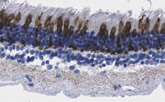
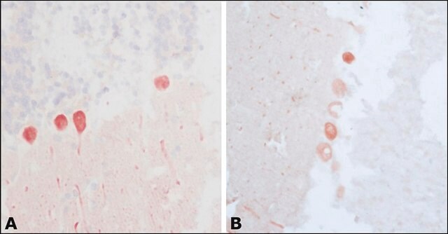
Anti-Opsin Red/Green, Cat. No. AB5405, is a rabbit polyclonal antibody that detects Opsin Red/Green and is tested for use in Immunohistochemistry (Paraffin).
Quality
Evaluated by Immunohistochemistry (Paraffin) in Mouse retina tissue sections.Immunohistochemistry (Paraffin) Analysis: A 1:250 dilution of this antibody detected Opsin Red/Green in Mouse retina tissue sections.
Physical form
Format: Purified
Purified rabbit polyclonal antibody in buffer containing 0.02 M phosphate buffer, pH 7.6, 0.25 M NaCl, and 0.1% sodium azide.
Storage and Stability
Recommended storage: +2°C to +8°C.
Other Notes
Concentration: Please refer to the Certificate of Analysis for the lot-specific concentration.
Legal Information
CHEMICON is a registered trademark of Merck KGaA, Darmstadt, Germany
Not finding the right product?
Try our Product Selector Tool.
recommended
Product No.
Description
Pricing
Storage Class Code
12 - Non Combustible Liquids
WGK
WGK 2
Flash Point(F)
Not applicable
Flash Point(C)
Not applicable
Certificates of Analysis (COA)
Search for Certificates of Analysis (COA) by entering the products Lot/Batch Number. Lot and Batch Numbers can be found on a product’s label following the words ‘Lot’ or ‘Batch’.
Already Own This Product?
Find documentation for the products that you have recently purchased in the Document Library.
Analysis of gene function in the retina.
Takahiko Matsuda,Constance L Cepko
Methods in Molecular Biology null
Rescue of retinal degeneration by intravitreally injected adult bone marrow-derived lineage-negative hematopoietic stem cells.
Otani, Atsushi, et al.
The Journal of Clinical Investigation, 114, 765-774 (2004)
Unique photoreceptor arrangements in a fish with polarized light discrimination.
Novales Flamarique I
The Journal of Comparative Neurology null
In vivo and in vitro development of S- and M-cones in rat retina.
Arango-Gonzalez, B; Szabo, A; Pinzon-Duarte, G; Lukats, A; Guenther, E; Kohler, K
Investigative Ophthalmology & Visual Science null
The electroretinogram (ERG) of a diurnal cone-rich laboratory rodent, the Nile grass rat (Arvicanthis niloticus).
Gilmour, Gregory S, et al.
Vision Research, 48, 2723-2731 (2008)
Our team of scientists has experience in all areas of research including Life Science, Material Science, Chemical Synthesis, Chromatography, Analytical and many others.
Contact Technical Service