
추천 제품


생물학적 소스
mouse
Quality Level
결합
unconjugated
항체 형태
purified immunoglobulin
항체 생산 유형
primary antibodies
클론
NNG-811, monoclonal
양식
buffered aqueous solution
분자량
~40 kDa
종 반응성
human
포장
antibody small pack of 25 μL
농도
~2 mg/mL
기술
immunocytochemistry: suitable
immunoprecipitation (IP): suitable
indirect ELISA: suitable
western blot: 4-8 μg/mL
동형
IgG1
UniProt 수납 번호
배송 상태
dry ice
저장 온도
−20°C
타겟 번역 후 변형
unmodified
유전자 정보
human ... NANOG(79923)
일반 설명
Anti-Nanog antibody, Mouse monoclonal (mouse IgG1 isotype) is derived from the hybridoma NNG-811 produced by the fusion of mouse myeloma cells and splenocytes from BALB/c mice immunized with recombinant human Nanog. The human Nanog gene product is a 305 amino acid, 35 kDa protein with a tryptophan repeat domain and two C-terminal trans-activating domains. It is primarily located in the nucleus.
Nanog is a homeodomain transcription factor expressed in cells of the inner cell mass of early embryos, in embryonic stem cells (ESC), germ cells and in stem/progenitor cells of some tissues. Together with Oct-4, Sox-2, and Rex-1, it is a molecular marker for pluripotent cells and for undifferentiated stem cells. This protein is crucial for the maintenance of pluripotency of ESCs and is down-regulated when ESCs differentiate.
Nanog controls the expression of many ESC genes together with other stem cell transcription factors like Oct-4 and Sox-2. Nanog targets both repressor and activator complexes to regulatory regions of hundreds of genes in the genome. Expression of nanog can be detected primarily in germ cell tumors and in tumors of other cell types. Nanog is an important marker for Seminomas, testicular carcinomas, teratocarcinomas, and germ cell-like tumors in various tissues. Furthermore, it was shown to transform NIH3T3 cells.
Nanog controls the expression of many ESC genes together with other stem cell transcription factors like Oct-4 and Sox-2. Nanog targets both repressor and activator complexes to regulatory regions of hundreds of genes in the genome. Expression of nanog can be detected primarily in germ cell tumors and in tumors of other cell types. Nanog is an important marker for Seminomas, testicular carcinomas, teratocarcinomas, and germ cell-like tumors in various tissues. Furthermore, it was shown to transform NIH3T3 cells.
특이성
Monoclonal Anti-Nanog reacts specifically with human Nanog.
면역원
recombinant human Nanog.
애플리케이션
Anti-Nanog antibody, Mouse monoclonal has been used in:
- enzyme-linked immunosorbent assay (ELISA)
- immunoblotting
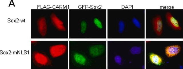
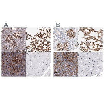
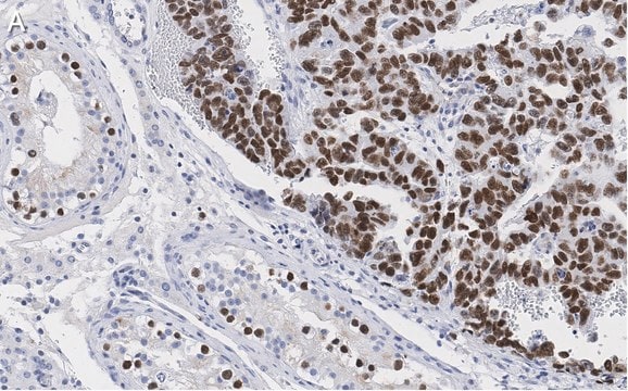
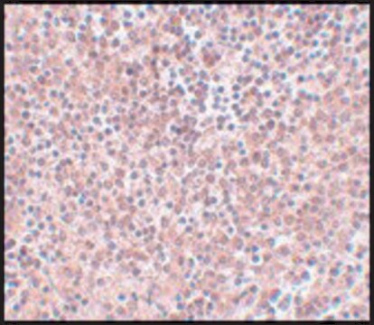
- immunocytochemistry
- flow cytometric analysis
- immunoprecipitation
- immunofluorescence
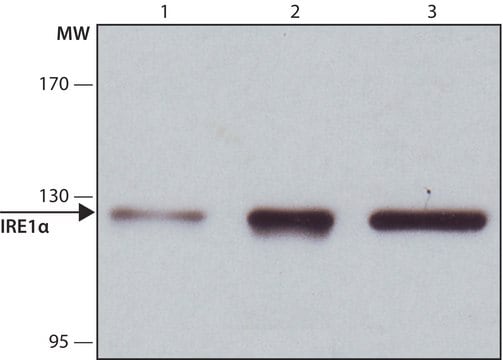
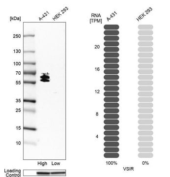
Monoclonal Anti-Nanog antibody is suitable for use in ELISA, immunoblotting (~ 40 kDa), immunocytochemistry and immunoprecipitation.
Immunoblotting: a working antibody concentration of 2-4 mg/mL is recommended using extracts of NT2 cells.
Immunoblotting: a working antibody concentration of 2-4 mg/mL is recommended using extracts of NT2 cells.
물리적 형태
Solution in 0.01 M phosphate buffered saline, pH 7.4, containing 15 mM sodium azide.
면책조항
Unless otherwise stated in our catalog or other company documentation accompanying the product(s), our products are intended for research use only and are not to be used for any other purpose, which includes but is not limited to, unauthorized commercial uses, in vitro diagnostic uses, ex vivo or in vivo therapeutic uses or any type of consumption or application to humans or animals.
적합한 제품을 찾을 수 없으신가요?
당사의 제품 선택기 도구.을(를) 시도해 보세요.
Storage Class Code
10 - Combustible liquids
WGK
WGK 3
Flash Point (°F)
Not applicable
Flash Point (°C)
Not applicable
개인 보호 장비
Eyeshields, Gloves, multi-purpose combination respirator cartridge (US)
The C-terminal pentapeptide of Nanog tryptophan repeat domain interacts with Nac1 and regulates stem cell proliferation but not pluripotency
Ma T, et al.
The Journal of Biological Chemistry, 284(24), 16071-16081 (2009)
NANOG reporter cell lines generated by gene targeting in human embryonic stem cells
Fischer Y, et al.
Testing, 5(9), e12533-e12533 (2010)
Anda Huna et al.
Cell cycle (Georgetown, Tex.), 14(18), 2969-2984 (2015-06-24)
Tumor cellular senescence induced by genotoxic treatments has recently been found to be paradoxically linked to the induction of "stemness." This observation is critical as it directly impinges upon the response of tumors to current chemo-radio-therapy treatment regimens. Previously, we
Jekaterina Erenpreisa et al.
Cell biology international, 35(7), 687-695 (2011-01-22)
'Neosis' describes the process whereby p53 function-deficient tumour cells undergo self-renewal after genotoxic damage apparently via senescing ETCs (endopolyploid tumour cells). We previously reported that autophagic digestion and extrusion of DNA occurs in ETC and subsequently revealed that self-renewal transcription
Marie Zbinden et al.
The EMBO journal, 29(15), 2659-2674 (2010-06-29)
A cohort of genes associated with embryonic stem (ES) cell behaviour, including NANOG, are expressed in a number of human cancers. They form an ES-like signature we first described in glioblastoma multiforme (GBM), a highly invasive and incurable brain tumour.
자사의 과학자팀은 생명 과학, 재료 과학, 화학 합성, 크로마토그래피, 분석 및 기타 많은 영역을 포함한 모든 과학 분야에 경험이 있습니다..
고객지원팀으로 연락바랍니다.