
추천 제품


생물학적 소스
rabbit
Quality Level
결합
unconjugated
항체 형태
IgG fraction of antiserum
항체 생산 유형
primary antibodies
클론
polyclonal
양식
buffered aqueous solution
분자량
antigen 40-42 kDa
종 반응성
human
기술
immunoprecipitation (IP): 100-150 μg using lysate of mitochondria from 2.5 to 5.0 × 10⁷ HeLa cells
microarray: suitable
western blot: 1:8,000 using a HeLa cell (human epithelioid carcinoma) mitochondria extract
UniProt 수납 번호
배송 상태
dry ice
저장 온도
−20°C
타겟 번역 후 변형
unmodified
유전자 정보
human ... CLEC4D(338339)
mouse ... Clec4d(17474)
rat ... Clec4d(362432)
일반 설명
Anti-Mcl-1 is developed in rabbit using a synthetic peptide corresponding to an internal region of Mcl-1 of human origin with N-terminal added cysteine, conjugated to maleimide activated keyhole limpet hemocyanin (KLH), as immunogen. MCL1 apoptosis regulator, BCL2 family member (Mcl-1) is expressed in many normal and neoplastic cells and is especially abundant in skeletal and cardiac muscle and in germinal centers of lymphoid tissues.
Mcl-1 is a member of Bcl-2 family that contains 3 Bcl-2 homology (BH) domains, BH1, BH2 and BH3, originally identified as an upregulated gene in human myeloid leukemia cell line (ML-1) in response to PMA. It is regulated transcriptionally and post-transcriptionally to enhance the cell survival. Mcl-1 is induced rapidly through cytokine-mediated survival pathways. Additionally, the upstream half of Mcl-1 RNA contains PEST (pro (P) Glu (E), Ser (S) and Thr (T) ) sequences and exhibits rapid turn-over. Increased expression of Mcl-1 maintains cell viability, decreased expression promotes cell death. Mcl-1 is reported to exhibit differentiation stage-specific expression in hematopoietic lineages and epithelial cells. A splice variant of Mcl-1, Mcl-1s promotes cell death. Often, the expression of Mcl-1 is induced via the MAPK signalling pathway acting on SRF/Elk-1 or Akt/CREB regulated pathway. Mcl-1 is expressed in many normal and neoplastic cells and is especially abundant in skeletal and cardiac muscle and in germinal centers of lymphoid tissues. It is predominantly expressed in the mitochondria but in neutrophils it seems to be mainly located in nuclear fractions.
특이성
Anti-Mcl-1 specifically recognizes Mcl-1 in tissue and cell extracts (40 to 42 kDa doublet).
면역원
synthetic peptide corresponding to an internal region of Mcl-1 of human origin (amino acids 121-139). This sequence is highly similar in mouse and rat.
애플리케이션
A minimum working dilution of 1:8000 is determined by immunoblotting using HeLa human epithelioid carcinoma mitochondria extract. It may also be used for detection by immunoblotting in Human melanoma cell lines, Human colon adenocarcinoma HT-29 and SW620 cells. Mcl-1 is immunoprecipitated from the lysate of mitochondria from 2.5 to 5.0x10 7 HeLa cells using 100 to 150 μg of the antibody. The antibody is suitable for protein microarray applications.
Anti-Mcl-1 antibody produced in rabbit has been used in immunoblotting.
생화학적/생리학적 작용
MCL1 apoptosis regulator, BCL2 family member (Mcl-1) exhibits great lability presumably due to its PEST sequence (P, Pro; E, Glu; S, Ser; T, Thr). Unlike the stable Bcl-2 protein, Mcl-1 exhibits great lability presumably due to its PEST sequence (P, Pro; E, Glu; S, Ser; T, Thr). Mcl-1, like Bcl-2, promotes cell viability under conditions which otherwise cause apoptosis. MCL1 acts as a chaperone of fortilin by binding and stabilizing it. It also interacts and negatively regulates the proliferating cell nuclear antigen (PCNA).
물리적 형태
Solution in 0.01 M phosphate buffered saline, pH 7.4, containing 15 mM sodium azide
면책조항
Unless otherwise stated in our catalog or other company documentation accompanying the product(s), our products are intended for research use only and are not to be used for any other purpose, which includes but is not limited to, unauthorized commercial uses, in vitro diagnostic uses, ex vivo or in vivo therapeutic uses or any type of consumption or application to humans or animals.
적합한 제품을 찾을 수 없으신가요?
당사의 제품 선택기 도구.을(를) 시도해 보세요.

신호어
Warning
유해 및 위험 성명서
Hazard Classifications
Acute Tox. 4 Dermal - Acute Tox. 4 Oral - Aquatic Chronic 3
Storage Class Code
10 - Combustible liquids
WGK
WGK 3
Flash Point (°F)
Not applicable
Flash Point (°C)
Not applicable
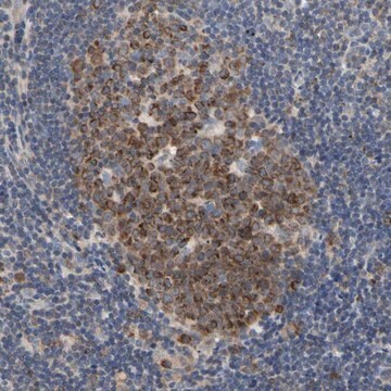
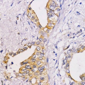
Immunohistochemical analysis of Mcl-1 protein in human tissues. Differential regulation of Mcl-1 and Bcl-2 protein production suggests a unique role for Mcl-1 in control of programmed cell death in vivo.
Krajewski S, et al.
The American Journal of Pathology, 146(6), 1309-1309 (1995)
Different modulation of TRAIL-induced apoptosis by inhibition of pro-survival pathways in TRAIL-sensitive and TRAIL-resistant colon cancer cells
Vaculova A, et al.
Febs Letters, 580(28-29), 6565-6569 (2006)
Differences in TRAIL-induced changes of Mcl-1 expression among distinct human colon epithelial cell lines
Vaculova A, et al.
Experimental Cell Research, 315(19), 3259-3266 (2009)
S Krajewski et al.
The American journal of pathology, 146(6), 1309-1319 (1995-06-01)
The mcl-1 gene encodes an approximately 37-kd protein that has significant homology with Bcl-2, an inhibitor of programmed cell death that is expressed in many types of long-lived cells. In this study we determined the in vivo patterns of Mcl-1
Doxorubicin and etoposide sensitize small cell lung carcinoma cells expressing caspase-8 to TRAIL
Vaculova A, et al.
Molecular Cancer, 9(1), 87-87 (2010)
자사의 과학자팀은 생명 과학, 재료 과학, 화학 합성, 크로마토그래피, 분석 및 기타 많은 영역을 포함한 모든 과학 분야에 경험이 있습니다..
고객지원팀으로 연락바랍니다.