
추천 제품


생물학적 소스
rabbit
Quality Level
결합
unconjugated
항체 형태
affinity isolated antibody
항체 생산 유형
primary antibodies
클론
polyclonal
제품 라인
Prestige Antibodies® Powered by Atlas Antibodies
양식
buffered aqueous glycerol solution
종 반응성
human
향상된 검증
orthogonal RNAseq
Learn more about Antibody Enhanced Validation
기술
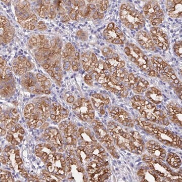
immunohistochemistry: 1:500- 1:1000
면역원 서열
HKRGMKGDTVNVRRSVRVKTKNPPHCLEITPPSSEKLVSVMRLSDLSTEDDDSGHCKMNRYDKKIDSLMNAVGCLKSEVKMQKGERQMAKRFLEERKEELEEVAHELAETEHENTVLRHNIERMKEEKDFTILQKKHLQQEKE
UniProt 수납 번호
배송 상태
wet ice
저장 온도
−20°C
타겟 번역 후 변형
unmodified
유전자 정보
human ... ODF2(4957)
일반 설명
The outer dense fiber of sperm tails 2 (ODF2) gene is located on the human chromosome at 9q34.11. This gene encodes for two proteins, namely testis-specific structural protein ODF2 and ubiquitous centriolar protein cenexin.
면역원
outer dense fiber of sperm tails 2 isoform 1 recombinant protein epitope signature tag (PrEST)
애플리케이션
Anti-ODF2 antibody produced in rabbit has been used in immunoblotting and immunofluorescence.
Anti-ODF2 antibody produced in rabbit, a Prestige Antibody, is developed and validated by the Human Protein Atlas (HPA) project . Each antibody is tested by immunohistochemistry against hundreds of normal and disease tissues. These images can be viewed on the Human Protein Atlas (HPA) site by clicking on the Image Gallery link. The antibodies are also tested using immunofluorescence and western blotting. To view these protocols and other useful information about Prestige Antibodies and the HPA, visit sigma.com/prestige.
생화학적/생리학적 작용
ODFs (outer dense fibres) consist of nine fibres surrounding the axoneme. They form the principal cytoskeletal structure of sperm tail. ODF2 is located at the sperm tail midpiece and principal piece. It has a thin cortex surrounded by central medulla. It is also involved in maintaining passive elastic structure and elastic recoil of the sperm tail and in protection of tail against shearing forces encountered during epididymal transport as well as during ejaculation.
특징 및 장점
Prestige Antibodies® are highly characterized and extensively validated antibodies with the added benefit of all available characterization data for each target being accessible via the Human Protein Atlas portal linked just below the product name at the top of this page. The uniqueness and low cross-reactivity of the Prestige Antibodies® to other proteins are due to a thorough selection of antigen regions, affinity purification, and stringent selection. Prestige antigen controls are available for every corresponding Prestige Antibody and can be found in the linkage section.
Every Prestige Antibody is tested in the following ways:
Every Prestige Antibody is tested in the following ways:
- IHC tissue array of 44 normal human tissues and 20 of the most common cancer type tissues.
- Protein array of 364 human recombinant protein fragments.
결합
Corresponding Antigen APREST85163
물리적 형태
Solution in phosphate-buffered saline, pH 7.2, containing 40% glycerol and 0.02% sodium azide
법적 정보
Prestige Antibodies is a registered trademark of Merck KGaA, Darmstadt, Germany
면책조항
Unless otherwise stated in our catalog or other company documentation accompanying the product(s), our products are intended for research use only and are not to be used for any other purpose, which includes but is not limited to, unauthorized commercial uses, in vitro diagnostic uses, ex vivo or in vivo therapeutic uses or any type of consumption or application to humans or animals.
적합한 제품을 찾을 수 없으신가요?
당사의 제품 선택기 도구.을(를) 시도해 보세요.
Storage Class Code
10 - Combustible liquids
WGK
WGK 1
Flash Point (°F)
Not applicable
Flash Point (°C)
Not applicable
개인 보호 장비
Eyeshields, Gloves, multi-purpose combination respirator cartridge (US)
Gregory Mazo et al.
Developmental cell, 39(4), 424-437 (2016-11-08)
Vertebrate cells can initiate ciliogenesis from centrioles at the cell center, near the Golgi, forming primary cilia confined or submerged in a deep narrow pit created by membrane invagination. How or why cells maintain submerged cilia is unclear. Here, by
Miho Ibi et al.
Journal of cell science, 124(Pt 6), 857-864 (2011-02-18)
The keratin cytoskeleton performs several functions in epithelial cells and provides regulated interaction sites for scaffold proteins, including trichoplein. Previously, we found that trichoplein was localized on keratin intermediate filaments and desmosomes in well-differentiated, non-dividing epithelia. Here, we report that
C Petersen et al.
Molecular human reproduction, 5(7), 627-635 (1999-06-25)
The outer dense fibres (ODF) are a main cytoskeletal structure of the sperm tail. Despite their importance in the morphology and function of the sperm tail, their constituents are poorly described. Here we investigate the protein composition of human outer
X Shao et al.
The Journal of biological chemistry, 272(10), 6105-6113 (1997-03-07)
The study of mammalian sperm tail outer dense fibers (ODF), a structure of unknown function, is hampered by the insoluble nature of ODF proteins and the availability of only one cloned component, Odf27. We report here the first use of
David Asante et al.
Journal of cell science, 126(Pt 22), 5189-5197 (2013-09-21)
The correct formation of primary cilia is central to the development and function of nearly all cells and tissues. Cilia grow from the mother centriole by extension of a microtubule core, the axoneme, which is then surrounded with a specialized
자사의 과학자팀은 생명 과학, 재료 과학, 화학 합성, 크로마토그래피, 분석 및 기타 많은 영역을 포함한 모든 과학 분야에 경험이 있습니다..
고객지원팀으로 연락바랍니다.