
추천 제품


일반 설명
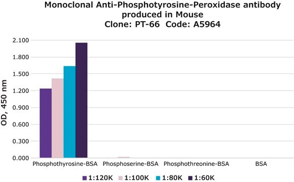
As determined by ELISA and competitive ELISA, the antibody reacts specifically with phosphorylated tyrosine, both as free amino acid or conjugated to carriers such as BSA or KLH. No cross-reactivity is observed with non-phosphorylated tyrosine, phosphothreonine, phosphoserine, AMP or ATP.
Monoclonal Anti-Phosphotyrosine (mouse IgG1 isotype) is derived from the hybridoma produced by the fusion of mouse myeloma cells and splenocytes from an immunized mouse.
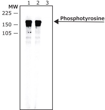
Reversible phosphorylation of proteins is an important post-translational modification that plays a regulatory role in the expression of most proteins in the cells. Reversible phosphorylation at multiple serine, tyrosine and threonine residues mediates numerous signalling pathways in both prokaryotic and eukaryotic cells . Cellular proteins with phosphorylated tyrosine increase many fold by the activation of tyrosine kinases. Most mitogenic receptor systems such as EGF, PDGF, insulin receptors contain serine/threonine/tyrosine kinase domains that undergo autophosphorylation when receptors bind to the respective ligands. Monoclonal anti-phosphotyrosine?biotin antibody can be used in dot blot (diluted 1:32,000). Mouse anti-phosphotyrosine?biotin antibody reacts specifically with phosphorylated tyrosine both as the free amino acid or when conjugated to BSA or KLH. This product does not react with non-phosphorylated tyrosine or other phosphorylated amino acids, including serine and threonine, or with phosphorylated molecules like AMP or ATP.
면역원
phosphotyrosine conjugated to BSA
애플리케이션
Monoclonal Anti-Phosphotyrosine?Biotin antibody produced in mouse has been used in western blot analysis to detect tyrosine phosphorylated proteins. It has also been used in BIAcore analysis. This conjugate maybe used as an analytical tool by enabling the identification and quantification of tyrosine phosphorylated proteins.
물리적 형태
Solution in 0.01 M phosphate buffered saline, pH 7.4, containing 1% bovine serum albumin and 15 mM sodium azide.
면책조항
Unless otherwise stated in our catalog or other company documentation accompanying the product(s), our products are intended for research use only and are not to be used for any other purpose, which includes but is not limited to, unauthorized commercial uses, in vitro diagnostic uses, ex vivo or in vivo therapeutic uses or any type of consumption or application to humans or animals.
적합한 제품을 찾을 수 없으신가요?
당사의 제품 선택기 도구.을(를) 시도해 보세요.
Storage Class Code
10 - Combustible liquids
WGK
nwg
Flash Point (°F)
Not applicable
Flash Point (°C)
Not applicable
Essential roles for Dok2 and RasGAP in CD200 receptor-mediated regulation of human myeloid cells
Mihrshahi R, et al.
Journal of Immunology, 183(8), 4879-4886 (2009)
Qiao Yan et al.
The Journal of neuroscience : the official journal of the Society for Neuroscience, 23(20), 7504-7509 (2003-08-22)
Alzheimer's disease (AD) is characterized by a microglial-mediated inflammatory response elicited by extensive amyloid deposition in the brain. Nonsteroidal anti-inflammatory drug (NSAID) treatment reduces AD risk, slows disease progression, and reduces microglial activation; however, the basis of these effects is
Senarath Dissanayake et al.
Immunology, 107(4), 411-419 (2002-12-04)
T helper type 2 (Th2) -polarized immune responses are characteristically dominant in helminth infections. Two murine models that show a Th1 to Th2 polarization with infection progression are those of Schistosoma mansoni and Taenia crassiceps. In both, an early Th1
Induction of immunoglobulin G1, interleukin-6 and interleukin-10 by Taenia crassiceps metacestode carbohydrates
Dissanayake S, et al.
Immunology, 107(4), 411-419 (2002)
J M Revest et al.
The European journal of neuroscience, 11(4), 1134-1147 (1999-04-02)
F3, a mouse glycosyl-phosphatidylinositol anchored molecule of the immunoglobulin superfamily, is known to influence axonal growth and fasciculation via multiple interactions of its modular immunoglobulin-like domains. We prepared an Fc chimeric molecule (F3IgFc) to identify molecules interacting with these domains
자사의 과학자팀은 생명 과학, 재료 과학, 화학 합성, 크로마토그래피, 분석 및 기타 많은 영역을 포함한 모든 과학 분야에 경험이 있습니다..
고객지원팀으로 연락바랍니다.