
추천 제품


사용
sufficient for ≤96 tests
Quality Level
포장
kit of 1 (9 components)
제조업체/상표
Roche
기술
ELISA: suitable
저장 온도
2-8°C
관련 카테고리
일반 설명
The Cell Death Detection ELISA serves as photometric enzyme immunoassay for the qualitative and quantitative in vitro determination of cytoplasmic histone-associated DNA fragments (mono- and oligonucleosomes) after induced cell death. This assay is based on the quantitative sandwich-enzyme immunoassay-principle using mouse monoclonal antibodies directed against DNA and histones, respectively. The antibodies used are not species specific.
특이성
Anti-histone antibody reacts with the histones H1, H2A, H2B, H3, and H4 of various species (e.g., human, mouse, rat, hamster, cow, opossum, and Xenopus). Anti-DNA-POD antibody binds to ss- and dsDNA. Therefore, the ELISA allows the detection of mono- and oligonucleosomes from various species, and may be applied to measure apoptotic cell death in many different cell systems.
애플리케이션
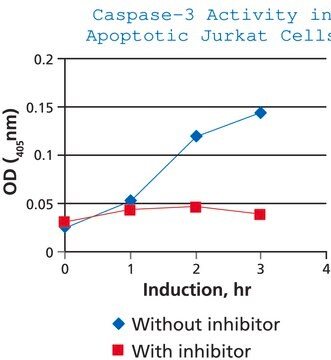
Cell Death Detection ELISA has been used to measure Caspase-3 activity, intracellular reactive oxygen species (ROS) and cell apoptosis.
For research use only. Not for use in diagnostic procedures.
Specific determination of mono- and oligonucleosomes in the cytoplasmic fraction of cell lysates.
Specific determination of mono- and oligonucleosomes in the cytoplasmic fraction of cell lysates.
포장
1 kit containing 9 components.
제조 메모
Working solution: Coating solution
Predilute 1 ml Coating buffer conc. with 9 ml double dist. water. Shortly before use, dilute 1 ml anti-histone antibody (reconstituted) with 9 ml Coating buffer.
Washing solution
Warm the Washing buffer concentrate to 15 to 25 °C and dilute 40 ml in 360 ml double distilled water. Mix thoroughly.
Sample solution
Preparation of the sample solution depends on the cell system used and the extent of cell death.
Example: Dilute 25 μl of sample in 225 μl Incubation buffer.
Conjugate solution
Dilute 1 ml Anti-DNA-POD (reconstituted) with 9 ml Incubation buffer.
Substrate solution
Depending on the number of samples tested, dissolve 1, 2, or 3 tablets in 5, 10, or 15 ml Substrate buffer.
Allow to come to 15 to 25 °C before use.
Note: The ABTS solution is light sensitive over extended periods of time.
Storage conditions (working solution): Anti-histone
2 to 8 °C for 2 months
Anti-DNA-POD
2 to 8 °C for 2 months
Coating solution
Prepare immediately before use!
Washing solution
2 to 8 °C for 2 months
Sample solution
2 to 8 °C for 2 months
Conjugate solution
Prepare immediately before use!
Substrate solution
2 to 8 °C for 1 month, protected from light.
재구성
Anti-histone
Reconstitute the lyophilizate in 1 ml double-distilled water for 10 minutes.Mix thoroughly.
Anti-DNA-POD
Reconstitute the lyophilizate in 1 ml double-distilled water for 10 minutes. Mix thoroughly.
Reconstitute the lyophilizate in 1 ml double-distilled water for 10 minutes.Mix thoroughly.
Anti-DNA-POD
Reconstitute the lyophilizate in 1 ml double-distilled water for 10 minutes. Mix thoroughly.
기타 정보
For life science research only. Not for use in diagnostic procedures.
키트 구성품 전용
제품 번호
설명
- Anti-histone antibody (clone H11–4)
- Anti-DNA-POD antibody (clone MCA-33)
- Coating Buffer
- Washing Buffer
- Incubation Buffer, ready-to-use solution
- Substrate Buffer, ready-to-use solution
- ABTS Substrate Tablet
- Microplate Modules
- Adhesive Plate Cover Foils
모두 보기 (9)


신호어
Danger
유해 및 위험 성명서
Hazard Classifications
Eye Dam. 1 - Skin Sens. 1
Storage Class Code
12 - Non Combustible Liquids
WGK
WGK 3
Flash Point (°F)
does not flash
Flash Point (°C)
does not flash
이미 열람한 고객
Gaya Hettiarachchi et al.
Molecular pharmaceutics, 13(3), 809-818 (2016-01-13)
Approximately, 40-70% of active pharmaceutical ingredients (API) are severely limited by their extremely poor aqueous solubility, and consequently, there is a high demand for excipients that can be used to formulate clinically relevant doses of these drug candidates. Here, proof-of-concept
Nilkantha Sen et al.
Nature cell biology, 10(7), 866-873 (2008-06-17)
Besides its role in glycolysis, glyceraldehyde-3-phosphate dehydrogenase (GAPDH) initiates a cell death cascade. Diverse apoptotic stimuli activate inducible nitric oxide synthase (iNOS) or neuronal NOS (nNOS), with the generated nitric oxide (NO) S-nitrosylating GAPDH, abolishing its catalytic activity and conferring
Silvia Di Loreto et al.
The international journal of biochemistry & cell biology, 40(2), 245-257 (2007-09-18)
The hippocampus is known to play a crucial role in learning and memory. Recent data from literature show that cognitive problems, common to aged or diabetic patients, may be related to accumulation of toxic alpha-oxoaldehydes such as methylglyoxal. Thus, it
Shikai Wang et al.
Scientific reports, 8(1), 17788-17788 (2018-12-14)
A growing number of studies have recently revealed a potential role for neutrophil extracellular traps (NETs) in the development of inflammation, coagulation and cell death. Deleterious consequences of NETs have been identified in ischemia-reperfusion (I/R)-induced organ damage, thrombosis and sepsis.
J P Heiserman et al.
Cell stress & chaperones, 20(3), 527-535 (2015-02-27)
Extracellular (ex) HSP60 is increasingly recognized as an agent of cell injury. Previously, we reported that low endotoxin exHSP60 causes cardiac myocyte apoptosis. Our findings supported a role for Toll-like receptor (TLR) 4 in HSP60 mediated apoptosis. To further investigate
자사의 과학자팀은 생명 과학, 재료 과학, 화학 합성, 크로마토그래피, 분석 및 기타 많은 영역을 포함한 모든 과학 분야에 경험이 있습니다..
고객지원팀으로 연락바랍니다.