
OP46
Anti-MDM2 (Ab-1) Mouse mAb (IF2)
liquid, clone IF2, Calbiochem®
동의어(들):
Anti-Murine Double Minute Chromosome-2, Anti-Ubiquitin Protein Ligase, Anti-p53 Binding Protein, Anti-Ubiquitin Protein Ligase, Anti-p53 Binding Protein, Anti-Murine Double Minute Chromosome-2
로그인조직 및 계약 가격 보기
모든 사진(1)
About This Item
UNSPSC 코드:
12352203
NACRES:
NA.41
추천 제품


생물학적 소스
mouse
Quality Level
항체 형태
purified antibody
항체 생산 유형
primary antibodies
클론
IF2, monoclonal
양식
liquid
포함
≤0.1% sodium azide as preservative (100 μg only)
종 반응성
human
반응하면 안 됨
mouse
제조업체/상표
Calbiochem®
저장 조건
do not freeze
동형
IgG2b
배송 상태
wet ice
저장 온도
2-8°C
타겟 번역 후 변형
unmodified
유전자 정보
human ... MDM2(4193)
mouse ... Mdm2(17246)
일반 설명
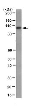
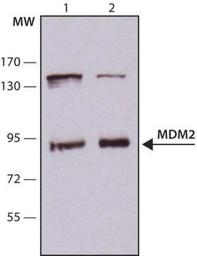
Purified mouse monoclonal antibody (see application references). Recognizes the ~90 kDa (apparent MW) MDM2 protein. Also recognizes isoforms at ~57 and ~74/76 kDa.
Recognizes the ~90 kDa (apparent MW) MDM2 protein. Also recognizes isoforms of ~57 kDa and ~74/76 kDa by immunoblotting.
This Anti-MDM2 (Ab-1) Mouse mAb (IF2) is validated for use in Frozen Sections Immunoblotting Immunofluorescence Immunoprecipitation Paraffin Sections for the detection of MDM2 (Ab-1).
면역원
Epitope: within amino acids 26-169 of human MDM2
Human
human MDM2
애플리케이션
Frozen Sections (1-5 µg/ml, see application references)
Immunoblotting (0.5-2 µg/ml, chemiluminescence)
Immunofluorescence (1-5 µg/ml)
Immunoprecipitation (1 µg/sample)
Paraffin Sections (1-5 µg/ml, heat pre-treatment required, see application references)
Immunoblotting (0.5-2 µg/ml, chemiluminescence)
Immunofluorescence (1-5 µg/ml)
Immunoprecipitation (1 µg/sample)
Paraffin Sections (1-5 µg/ml, heat pre-treatment required, see application references)
포장
Please refer to vial label for lot-specific concentration.
경고
Toxicity: Standard Handling (A)
물리적 형태
In 50 mM sodium phosphate buffer, 0.2% gelatin.
분석 메모
Positive Control
OSA-CL cells
OSA-CL cells
기타 정보
Although the amino acid sequence of MDM2 predicts a protein with a molecular mass of approximately 54 kDa, MDM2 protein migrates on SDS/PAGE with an apparent mobility of 90 kDa.
Immunoblotting Protocol
MDM2 (Ab-1) can be used to detect MDM2 by Western blot of proteins previously separated by SDS/PAGE and electrophoretically transferred onto nitrocellulose membranes. The proteins are reacted with the monoclonal antibody and visualized using an HRP conjugated goat anti-mouse antibody with chemiluminescent detection.
Materials
Equipment:
• Electrophoresis apparatus
• Electroblotting apparatus
• Rocker platform
Solutions and Reagents
• Anti-MDM2 (Ab-1) Mouse mAb (IF2) Cat. No. OP46 or OP46T
• HRP conjugated goat anti-mouse IgG heavy and light chains (e.g. Cat. No. 401215)
• Chemiluminescence detection system
• ELB Buffer (include a cocktail of proetease inhibitors, such as 0.5 µg/ml leupeptin, 1 µg/ml pepstatin, 1 mM EDTA and 0.2 mM PMSF): 50 mM Hepes pH 7.0, 250 mM NaCl, 0.5 mM EDTA, 0.1% Nonidet P-40 Alternative
• SDS-PAGE (7% acrylamide)
• Phosphate buffered saline (PBS) pH 7.4; 1 Liter: 0.2 g KCl, 0.2 g KH2PO4, 8 g NaCl, 1.15 g Na2HPO4
• PBS/0.1% Tween®-20 detergent (PBST)
• 3% Non-fat Dry Milk in PBST
Procedure
1. Lyse cells in ELB Buffer. (Alternatively, cells can be lysed in RIPA Buffer or directly into 1x Laemmli Sample Buffer).
2. Electrophorese 50-100 µg lysate using a 7% acrylamide gel.
3. Transfer the protein samples from the polyacrylamide gel onto a nitrocellulose membrane using an electroblotting apparatus.
4. Block the membrane for 1 h in PBST containing 3% non-fat dry milk at room temperature with rocking. Use about 1 ml per cm2 of membrane.
5. Incubate the membrane with 1 µg/ml Anti-MDM2 (Ab-1) Mouse mAb (IF2) in 3% non-fat dry milk/ PBST for 1 h at room temperature with rocking.
6. Wash the membrane 3 times, 15 min each, in PBST at room temperature with rocking.
7. Incubate the membrane with HRP conjugated goat anti-mouse IgG heavy and light chain antibody, diluted according to the supplier’s instructions, in 3% non-fat dry milk/ PBST at room temperature for 1 h.
8. Wash the membrane 4 times, 15 min each, in PBST at room temperature with rocking.
9. Develop the membrane using chemiluminescent detection reagents according to manufacturer instructions.
10. Expose the membrane to film for ten minutes. Adjust subsequent exposure times as needed.
Gorgoulis, V.G., et al. 1996. J. Pathol.180, 129.
Marchetti, A., et al. 1995. J. Pathol.175, 31.
Barak, Y., et al. 1993. EMBO J.12, 461.
Ladanyi, M., et al. 1993. Cancer Res.53, 16.
Leach, F.S., et al. 1993. Cancer Res.53, 2231.
Oliner, J.D., et al. 1993. Nature362, 857.
Momand, J., et al. 1992. Cell69, 1237.
Oliner, J.D., et al. 1992. Nature358, 80.
Fakharzadeh, S.S., et al. 1991. EMBO J. 10, 1565.
법적 정보
CALBIOCHEM is a registered trademark of Merck KGaA, Darmstadt, Germany
TWEEN is a registered trademark of Croda International PLC
적합한 제품을 찾을 수 없으신가요?
당사의 제품 선택기 도구.을(를) 시도해 보세요.
Storage Class Code
10 - Combustible liquids
WGK
nwg
Flash Point (°F)
Not applicable
Flash Point (°C)
Not applicable
시험 성적서(COA)
제품의 로트/배치 번호를 입력하여 시험 성적서(COA)을 검색하십시오. 로트 및 배치 번호는 제품 라벨에 있는 ‘로트’ 또는 ‘배치’라는 용어 뒤에서 찾을 수 있습니다.
Yongliang Hu et al.
Oncogene, 38(5), 731-746 (2018-09-05)
Our previous studies revealed that GADD45α is a liable protein, which undergoes MDM2-dependent constitutive ubiquitination and degradation in resting HepG2 hepatoma cells. Arsenite exposure induces ribosomal stress responses mediated by the ribosomal protein S7, which can block MDM2 activity and
Deborah J Luessen et al.
The Journal of biological chemistry, 294(38), 14068-14080 (2019-08-02)
Acute alcohol exposure alters the trafficking and function of many G-protein-coupled receptors (GPCRs) that are associated with aberrant behavioral responses to alcohol. However, the molecular mechanisms underlying alcohol-induced changes in GPCR function remain unclear. β-Arrestin is a key player involved
C Wasylyk et al.
Molecular and cellular biology, 20(15), 5554-5570 (2000-07-13)
The cell cycle arrest and proapoptotic functions of p53 are under tight control by Mdm2. After stress activation of p53 by nontranscriptional mechanisms, transcription of the mdm2 gene results in increased synthesis of Mdm2 and down-regulation of p53. Disruption of
Jennifer Hüllein et al.
Cancer research, 79(12), 3125-3138 (2019-04-20)
Oncogenic MYC activation promotes proliferation in Burkitt lymphoma, but also induces cell-cycle arrest and apoptosis mediated by p53, a tumor suppressor that is mutated in 40% of Burkitt lymphoma cases. To identify molecular dependencies in Burkitt lymphoma, we performed RNAi-based
Stephen L Lessnick et al.
Cancer cell, 1(4), 393-401 (2002-06-28)
Ewing's sarcoma is associated with a fusion between the EWS and FLI1 genes, forming an EWS/FLI fusion protein. We developed a system for the identification of cooperative mutations in this tumor through expression of EWS/FLI in primary human fibroblasts. Gene
자사의 과학자팀은 생명 과학, 재료 과학, 화학 합성, 크로마토그래피, 분석 및 기타 많은 영역을 포함한 모든 과학 분야에 경험이 있습니다..
고객지원팀으로 연락바랍니다.