
추천 제품


생물학적 소스
mouse
Quality Level
항체 형태
purified immunoglobulin
항체 생산 유형
primary antibodies
클론
11-5B, monoclonal
종 반응성
canine, sheep, mouse, pig, bovine, rat, rabbit, human
반응하면 안 됨
guinea pig, chicken
제조업체/상표
Chemicon®
기술
immunocytochemistry: suitable
immunohistochemistry (formalin-fixed, paraffin-embedded sections): suitable
western blot: suitable
동형
IgG1
NCBI 수납 번호
UniProt 수납 번호
배송 상태
wet ice
타겟 번역 후 변형
unmodified
유전자 정보
human ... CNP(1267)
일반 설명
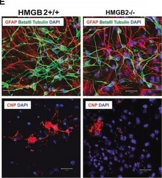
CNPase (2′, 3′-cyclic nucleotide 3′-phosphodiesterase [or -phosphohydrolase], EC 3.1.4.37) is present in very high levels in brain and peripheral nerve. This enzyme is found almost exclusively in oligo-dendrocytes and Schwann cells, the cells that form myelin in the central and peripheral nervous system, respectively.
Immunohistochemical localization of CNPase has shown the enzyme to be restricted to oligodendrocytes and Schwann cells. The enzyme appears to be distributed in single and double loose wraps of myelin and not in compact myelin as earlier thought by most investigators. CNPase is located on the inner and outer loops of myelin, paranodally and near the inner surface of the oligodendrocyte membrane. In plaque regions of multiple sclerosis patients, the enzyme is reduced, and when CNS myelin is decreased, CNPase is one of the earlier proteins to be lost from the myelin. In addition, an enzyme that is probably identical to brain CNPase is located in the outer rod segments within the visual system, and this protein is also recognized by the monoclonal antibody 11-5B. In mixed human gliomas, the enzyme levels are reduced, although about 5% of the oligodendrocytes occasionally show normal positive staining.
Immunohistochemical localization of CNPase has shown the enzyme to be restricted to oligodendrocytes and Schwann cells. The enzyme appears to be distributed in single and double loose wraps of myelin and not in compact myelin as earlier thought by most investigators. CNPase is located on the inner and outer loops of myelin, paranodally and near the inner surface of the oligodendrocyte membrane. In plaque regions of multiple sclerosis patients, the enzyme is reduced, and when CNS myelin is decreased, CNPase is one of the earlier proteins to be lost from the myelin. In addition, an enzyme that is probably identical to brain CNPase is located in the outer rod segments within the visual system, and this protein is also recognized by the monoclonal antibody 11-5B. In mixed human gliomas, the enzyme levels are reduced, although about 5% of the oligodendrocytes occasionally show normal positive staining.
특이성
CNPase, 48 and 46 kD polypeptides by SDS-PAGE. Differentiates clearly oligodendrocytes and Schwann cells from neurons, astrocytes, etc.
면역원
Purified human brain CNPase
애플리케이션
Detect CNPase using this Anti-CNPase Antibody, clone 11-5B validated for use in IC, IH, IH(P) & WB.
Immunocytochemistry:
A previous lot was used on primary oligodendrocyte cultures.
Immunohistochemistry:
A previous lot was used on rat hippocampus tissue and rat spinal cord.
Immunoblotting of myelin, the Wolfgram protein fraction, the SN4 fraction, tissue sections and mixed glial tumors (oligodendrogliomas, etc.) CNPase I (46 kDa) and CNPase II (48 kDa), which are differentially regulated during development, with the larger protein being expressed earlier than CNPase I during development.
Immunohistochemistry on both fresh frozen and paraffin embedded tissue (microwave pretreatment, ctirate pH 6.0).
Immunoblot:
Immunoblotting of myelin, the Wolfgram protein fraction, the SN4 fraction, tissue sections
and mixed glial tumors (oligodendrogliomas, etc.)
Optimal working dilutions must be determined by end user.
Research Category
Neuroscience
Neuroscience
Research Sub Category
Neuronal & Glial Markers
Neuronal & Glial Markers
품질
Routinely evaluated by Western Blot on Mouse Brain lysates.
Western Blot Analysis:
1:1000 dilution of this lot detected CNPASE on 10 μg of Mouse Brain lysates.
Western Blot Analysis:
1:1000 dilution of this lot detected CNPASE on 10 μg of Mouse Brain lysates.
표적 설명
48 & 46 kDa
물리적 형태
Format: Purified
Protein A purified
Purified mouse monoclonal IgG1 in buffer containing 0.02M phosphate buffer, 0.25 M NaCl with 0.1% sodium azide
저장 및 안정성
Stable for 1 year at 2-8ºC from date of receipt.
분석 메모
Control
Western Blot: Fresh bovine whole brain extract, mouse brain lysate.
Immunohistochemistry: Rat hippocampus tissue, rat spinal cord tissue.
Western Blot: Fresh bovine whole brain extract, mouse brain lysate.
Immunohistochemistry: Rat hippocampus tissue, rat spinal cord tissue.
기타 정보
Concentration: Please refer to the Certificate of Analysis for the lot-specific concentration.
법적 정보
CHEMICON is a registered trademark of Merck KGaA, Darmstadt, Germany
면책조항
Unless otherwise stated in our catalog or other company documentation accompanying the product(s), our products are intended for research use only and are not to be used for any other purpose, which includes but is not limited to, unauthorized commercial uses, in vitro diagnostic uses, ex vivo or in vivo therapeutic uses or any type of consumption or application to humans or animals.
적합한 제품을 찾을 수 없으신가요?
당사의 제품 선택기 도구.을(를) 시도해 보세요.
Storage Class Code
10 - Combustible liquids
WGK
WGK 2
시험 성적서(COA)
제품의 로트/배치 번호를 입력하여 시험 성적서(COA)을 검색하십시오. 로트 및 배치 번호는 제품 라벨에 있는 ‘로트’ 또는 ‘배치’라는 용어 뒤에서 찾을 수 있습니다.
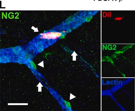
Electroconvulsive seizures induce proliferation of NG2-expressing glial cells in adult rat amygdala.
Malin Wennstrom, Johan Hellsten, Anders Tingstrom
Biological Psychiatry null
Hereditary cerebellar degenerative disease (cerebellar cortical abiotrophy) in rabbits.
Sato, J; Sasaki, S; Yamada, N; Tsuchitani, M
Veterinary Pathology null
S U Kim et al.
Brain research, 300(1), 195-199 (1984-05-21)
Previous biochemical studies have shown that high enzyme activity of 2':3'-cyclic-nucleotide 3'-phosphodiesterase (CNP) is found in isolated myelin and oligodendrocytes. We report here the specific and intense immunofluorescence staining of cultured oligodendrocytes obtained from the brains of mouse, rat, calf
H J Sheedlo et al.
Brain research, 288(1-2), 330-333 (1983-12-12)
The distribution of 2':3'-cyclic nucleotide 3'-phosphodiesterase (CNP) in fixed-tissue sections and Percoll-isolated cells (oligodendrocytes) of bovine cerebrum was studied by immunofluorescent techniques. Fluorescence in tissue sections exposed to CNP antisera was restricted to oligodendrocyte-like cells. Also, Percoll-isolated cells (oligodendrocytes) were
The Use of Interleukin 12-secreting Neural Stem Cells for the Treatment of Intracranial Glioma
Ehtesham, M., et al.
Cancer Research, 62, 5657-5663 (2002)
자사의 과학자팀은 생명 과학, 재료 과학, 화학 합성, 크로마토그래피, 분석 및 기타 많은 영역을 포함한 모든 과학 분야에 경험이 있습니다..
고객지원팀으로 연락바랍니다.