HPA001238
Anti-MMP9 antibody produced in rabbit

affinity isolated antibody, buffered aqueous glycerol solution
Synonym(s):
CLG4B
Sign Into View Organizational & Contract Pricing
Select a Size
All Photos(7)
Select a Size
Change View
About This Item
Recommended Products
biological source
rabbit
conjugate
unconjugated
antibody form
affinity isolated antibody
antibody product type
primary antibodies
clone
polyclonal
product line
Prestige Antibodies® Powered by Atlas Antibodies
form
buffered aqueous glycerol solution
species reactivity
human
enhanced validation
orthogonal RNAseq
Learn more about Antibody Enhanced Validation
technique(s)
immunofluorescence: 0.25-2 μg/mL
immunohistochemistry: 1:500-1:1000
General description
Matrix metalloproteinase-9 (MMP-9) is a pro-inflammatory, zinc-dependent proteinase, which is also known as 92kDa gelatinase. It is secreted by granulocytes and mononuclear cells. The activity of MMP-9 is controlled by gene transcription. The gene encoding it is localized on human chromosome 20q11.2-q13.1 and has 13 exons.
Immunogen
Recombinant protein corresponding to Matrix metalloproteinase-9
Sequence
PRQRQSTLVLFPGDLRTNLTDRQLAEEYLYRYGYTRVAEMRGESKSLGPALLLLQKQLSLPETGELDSATLKAMRTPRCGVPDLGRFQTFEGDLKWHHHNITYWIQNYSEDLPRAVIDDAFARAFALWSAVTPLTFTRVYSRDADI
Sequence
PRQRQSTLVLFPGDLRTNLTDRQLAEEYLYRYGYTRVAEMRGESKSLGPALLLLQKQLSLPETGELDSATLKAMRTPRCGVPDLGRFQTFEGDLKWHHHNITYWIQNYSEDLPRAVIDDAFARAFALWSAVTPLTFTRVYSRDADI
Application
All Prestige Antibodies Powered by Atlas Antibodies are developed and validated by the Human Protein Atlas (HPA) project and as a result, are supported by the most extensive characterization in the industry.
The Human Protein Atlas project can be subdivided into three efforts: Human Tissue Atlas, Cancer Atlas, and Human Cell Atlas. The antibodies that have been generated in support of the Tissue and Cancer Atlas projects have been tested by immunohistochemistry against hundreds of normal and disease tissues and through the recent efforts of the Human Cell Atlas project, many have been characterized by immunofluorescence to map the human proteome not only at the tissue level but now at the subcellular level. These images and the collection of this vast data set can be viewed on the Human Protein Atlas (HPA) site by clicking on the Image Gallery link. We also provide Prestige Antibodies® protocols and other useful information.
The Human Protein Atlas project can be subdivided into three efforts: Human Tissue Atlas, Cancer Atlas, and Human Cell Atlas. The antibodies that have been generated in support of the Tissue and Cancer Atlas projects have been tested by immunohistochemistry against hundreds of normal and disease tissues and through the recent efforts of the Human Cell Atlas project, many have been characterized by immunofluorescence to map the human proteome not only at the tissue level but now at the subcellular level. These images and the collection of this vast data set can be viewed on the Human Protein Atlas (HPA) site by clicking on the Image Gallery link. We also provide Prestige Antibodies® protocols and other useful information.
Anti-MMP9 antibody produced in rabbit has been used in:
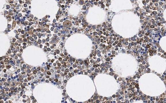
- immunohistochemistry
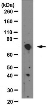
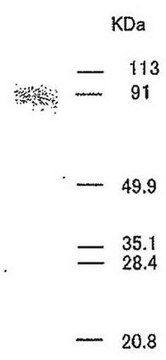
- western blotting
- fluorescence immunohistochemistry
Biochem/physiol Actions
Matrix metalloproteinase-9 (MMP-9) catalyzes the degradation of gelatin and collagen. It has been studied as a biomarker for inflammation in multiple sclerosis (MS). The MMP9 protein is a zinc-dependent protease which is responsible for degradation of extracellular matrix components. It also participates in the pathogenesis of cancers.
Features and Benefits
Prestige Antibodies® are highly characterized and extensively validated antibodies with the added benefit of all available characterization data for each target being accessible via the Human Protein Atlas portal linked just below the product name at the top of this page. The uniqueness and low cross-reactivity of the Prestige Antibodies® to other proteins are due to a thorough selection of antigen regions, affinity purification, and stringent selection. Prestige antigen controls are available for every corresponding Prestige Antibody and can be found in the linkage section.
Every Prestige Antibody is tested in the following ways:
Every Prestige Antibody is tested in the following ways:
- IHC tissue array of 44 normal human tissues and 20 of the most common cancer type tissues.
- Protein array of 364 human recombinant protein fragments.
Linkage
Corresponding Antigen APREST83039
Physical form
Solution in phosphate-buffered saline, pH 7.2, containing 40% glycerol and 0.02% sodium azide
Legal Information
Prestige Antibodies is a registered trademark of Merck KGaA, Darmstadt, Germany
Disclaimer
Unless otherwise stated in our catalog or other company documentation accompanying the product(s), our products are intended for research use only and are not to be used for any other purpose, which includes but is not limited to, unauthorized commercial uses, in vitro diagnostic uses, ex vivo or in vivo therapeutic uses or any type of consumption or application to humans or animals.
Not finding the right product?
Try our Product Selector Tool.
Storage Class Code
10 - Combustible liquids
WGK
WGK 1
Flash Point(F)
Not applicable
Flash Point(C)
Not applicable
Personal Protective Equipment
dust mask type N95 (US), Eyeshields, Gloves
Choose from one of the most recent versions:
Already Own This Product?
Find documentation for the products that you have recently purchased in the Document Library.
Julie A Bass et al.
BMC gastroenterology, 15, 129-129 (2015-10-16)
Early manifestations of pediatric inflammatory bowel disease (IBD) can be relatively nonspecific. Initial mucosal biopsies may not be conclusive, delaying the diagnosis until subsequent biopsies demonstrate typical histologic features of IBD. We hypothesized that certain inflammatory cell types may be
Miconazole protects blood vessels from MMP9-dependent rupture and hemorrhage.
Yang R, et al.
Disease models & mechanisms, 10(3), 337-348 (2017)
Matrix metalloproteinases: old dogs with new tricks.
Somerville RP, et al.
Genome Biology, 4(6), 216-216 (2003)
David G P van IJzendoorn et al.
Clinical cancer research : an official journal of the American Association for Cancer Research (2022-08-26)
A major component of cells in Tenosynovial Giant Cell Tumor (TGCT) consists of bystander macrophages responding to CSF1 that is overproduced by a small number of neoplastic cells with a chromosomal translocation involving the CSF1 gene. An autocrine loop was
Association between four MMP-9 polymorphisms and breast cancer risk: a meta-analysis.
Zhang X, et al.
Medical Science Monitor : International Medical Journal of Experimental and Clinical Research, 21(16), 1115-1115 (2015)
Our team of scientists has experience in all areas of research including Life Science, Material Science, Chemical Synthesis, Chromatography, Analytical and many others.
Contact Technical Service


